Your Cart Is Empty
✖No products in the cart.
Every implant begins with a fundamental challenge: accurately transferring the position of implants from the patient to the laboratory.
The final prosthesis can only be as accurate as the data used to design it. Even small errors can lead to misfit, extra chair time, component complications, and costly remakes.
Historically, clinicians relied on conventional impression techniques using splinted impression copings and physical models. While these methods have been widely used for decades, they introduce multiple opportunities for distortion throughout the workflow.
Digital dentistry improved this process significantly. Intraoral scanners eliminated many of the limitations associated with traditional impressions. But in full-arch implant cases, many clinicians still found one major limitation: precisely capturing implant positions across a long span.
This challenge led to the emergence of dental photogrammetry.
Today, photogrammetry is rapidly becoming one of the most important technologies in modern implant dentistry. It provides a highly precise measurement of implant positions across an entire arch.
Dental photogrammetry is a digital measurement technology that records the precise 3D position of dental implants using calibrated optical imaging.
Unlike traditional impressions or intraoral scanners, photogrammetry does not rebuild implant positions from surface data. It directly measures the spatial relationship between implants.
The technology is not new. Photogrammetry has been used for decades in industries like aerospace engineering, industrial metrology, and automotive manufacturing. These industries rely on it when accuracy must be measured in microns.
Photogrammetry for dental implants uses the same technology.
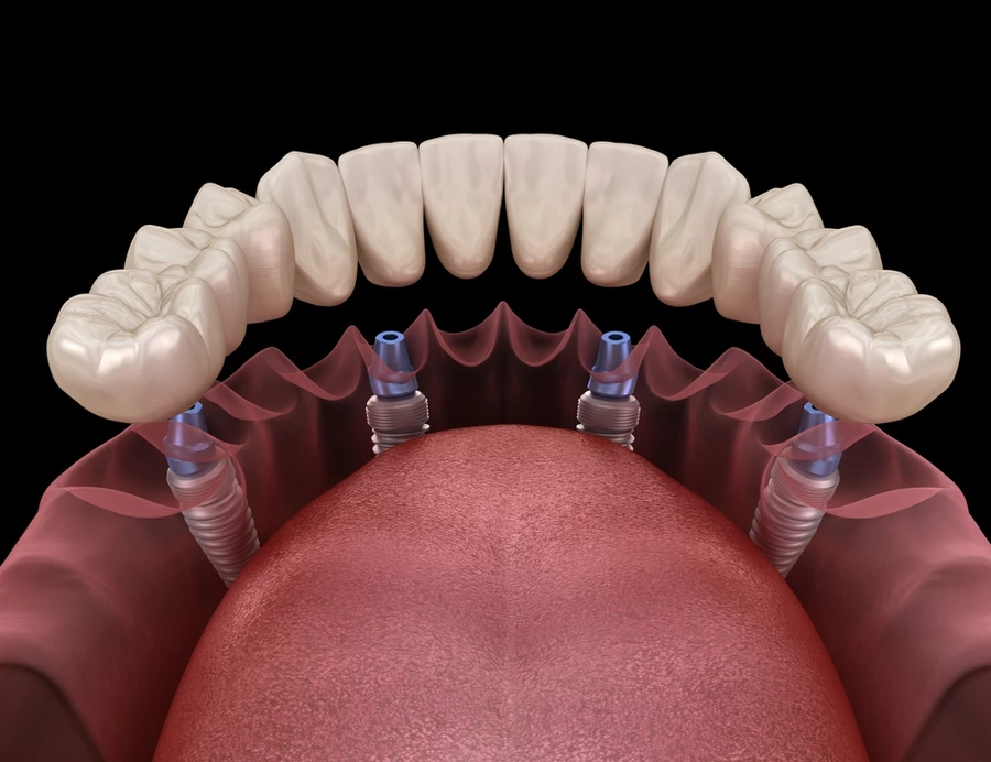
Specialized scanbodies are connected to implants or multi-unit abutments (MUAs). A photogrammetry device then captures multiple images from different angles. Sophisticated software then calculates the exact position of each implant in 3D space.
The result is a highly precise digital map of the implant sites. This data can then be used for prosthetic design and fabrication.
Precision is not just a technical detail. It directly affects fit, patient comfort, and long-term prosthetic success.
When implant positions are captured incorrectly, the final prosthesis may not seat passively. That can lead to:
The goal is always passive fit. This means the prosthesis seats precisely without placing harmful strain on the implants, abutments, or framework. Passive fit is especially important in full-arch cases.
In full-arch dentistry, the margin for error is small. Many clinicians refer to a working range of roughly 100–150 microns for clinically acceptable fit. For perspective, that is about the width of a human hair.
As the number of implants increases, this challenge becomes harder. A single implant may be more forgiving. Four, six, or more implants connected by one prosthesis are not.
This is where photogrammetry shows its greatest value.
Specialized photogrammetry scanbodies are attached to the implants or MUAs.
These scanbodies act as reference points. Their shape and dimensions are known, so the system can identify and measure them with precision.
The photogrammetry device captures multiple images of all scanbodies simultaneously.
The key difference is that photogrammetry records the relationship between implants directly. It does not rely on stitching together thousands of surface images across the arch.
This matters because stitching errors can compound as your anterior-posterior spread increases.
The software analyzes the images captured from multiple viewpoints. The system calculates:
The result is a precise digital record of the implant framework.
ICam photogrammetry runs 60,800 calculations, while other digital implant impression systems may run only 150–260 calculations. In a workflow where microns matter, more complete measurement data means more confidence.
Photogrammetry captures implant positions. But clinicians still require soft tissue anatomy and intraoral structures for prosthetic design. That is why photogrammetry is often combined with intraoral scanning.
The photogrammetry file provides the implant position data. The intraoral scan provides gingival contours, emergence profiles, occlusion, and surrounding anatomy.
Together, these files create a complete digital patient record.
Since it’s common in modern dentistry to use both intraoral scanning and photogrammetry, let’s talk about the differences. They are not competing technologies; they serve different purposes.
Intraoral scanners excel at capturing:
But intraoral scanners rely on image stitching. As the scan moves across a full arch, small alignment errors can build.
Photogrammetry does not estimate implant locations from surrounding surfaces. It measures them directly.
This becomes more important as the number of implants increases and the restoration becomes larger.
A 2026 systematic review found that photogrammetry systems showed significantly greater trueness and precision than intraoral scanners for capturing 3D implant positions in complete-arch and multi-implant restorations.
That is why many advanced full-arch workflows use both technologies: intraoral scanning for anatomy and photogrammetry for implant position.
If your practice is committed to accuracy, predictability, and better outcomes in full-arch implant dentistry, it may be time to rethink how implant positions are captured.
By directly measuring implant positions, rather than reconstructing them from impression materials or stitched surface images, dental photogrammetry offers a higher level of accuracy. Photogrammetry is transforming full-arch restorations and implant workflows around the world.
Discover how ICam system is helping clinicians move beyond estimation and toward true measurement.
No products in the cart.